Spine MRI in Seminole, FL
A spine MRI is the gold-standard scan for back and neck pain, sciatica, herniated discs, and spinal stenosis. No radiation, no referral needed for self-pay, and same-week scheduling at our Seminole, FL imaging center.
What Is a Spine MRI?
A spine MRI uses a strong magnetic field and radio waves — no radiation — to produce detailed cross-sectional images of your spine: the vertebrae, the discs between them, the spinal cord, and the surrounding nerves, muscles, and soft tissue. It is the single most accurate test for evaluating back and neck pain, because it shows soft-tissue structures (discs, nerves, the spinal cord) that an X-ray or CT simply cannot. A spine MRI can be focused on the neck (cervical spine), the mid-back (thoracic spine), or the lower back (lumbar spine), depending on where your symptoms are. Most spine MRIs are performed without contrast; contrast is added only when your physician needs to evaluate infection, tumor, or post-surgical changes.
What Can Spine MRI Diagnose?
- Herniated, bulging, or degenerated discs
- Sciatica and pinched / compressed nerves
- Spinal stenosis (narrowing of the spinal canal)
- Lower back pain and chronic neck pain causes
- Spinal cord injury, compression, and inflammation
- Arthritis and degenerative disc disease
- Spinal tumors and bone metastases
- Infection (discitis, osteomyelitis) and post-surgical changes
Spine MRI vs. X-Ray vs. CT for Back Pain
For most back and neck pain, MRI is the gold standard because it shows soft tissue — discs, nerves, and the spinal cord — that other scans miss.
| Feature | AMI ✓ | X-Ray | CT |
|---|---|---|---|
| Shows discs & nerves | Yes — detailed soft-tissue detail | No — bone only | Limited |
| Shows spinal cord | Yes | No | Limited |
| Radiation | None | Low dose | Higher dose |
| Best for | Herniated disc, sciatica, stenosis, nerve compression | Fractures, alignment | Bony detail, surgical planning |
| Self-pay from | $295 (single region) | $50 | From $295 |
How to Prepare for Your Spine MRI
- 1Remove all metal jewelry, piercings, watches, and hair clips
- 2Tell our technologist about any implants, pacemakers, spinal hardware, or surgical clips
- 3Wear comfortable clothing without metal zippers, buttons, or underwire
- 4Arrive 15 minutes early to complete intake and safety screening
- 5If using insurance, bring your insurance card and physician's order
- 6If you are anxious in enclosed spaces, ask about our Open MRI option
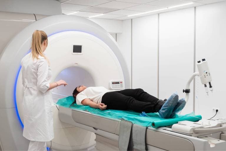
What to Expect During Your Spine MRI
Check-In & Safety Screening
You'll complete a safety questionnaire about any metal or implants in your body. Our technologist reviews your history and confirms which region of the spine is being imaged.
Positioning
You'll lie on your back on a padded table that slides into the MRI. A coil is placed over the area of your spine being scanned. We'll give you earplugs or headphones with music to make you comfortable.
The Scan
You'll hear rhythmic tapping and humming — completely normal. The key is to stay as still as possible so the images are sharp. A typical spine MRI takes 30–45 minutes depending on how many regions are imaged.
After Your Scan
You can drive, return to work, and resume all normal activities immediately. A board-certified radiologist reads your images and delivers results to your physician within 48 hours; stat reads are available for urgent cases.
Frequently Asked Questions
Our self-pay spine MRI starts at $295 for a single region without contrast — up to 75% less than typical hospital pricing. The exact price depends on how many regions (cervical, thoracic, lumbar) are imaged and whether contrast is needed. Call (727) 398-5999 for an exact quote, or see our transparent self-pay pricing page.
Self-pay patients do not need a referral. Our physician reviews your request the same day — most self-pay patients are seen same day or added as a walk-in. If you plan to use insurance, a physician's order is still required, and we will verify your benefits before your appointment. Call (727) 398-5999 and we'll get you scheduled.
Most spine MRI exams take 30–45 minutes. Scanning a single region (for example, the lumbar spine) is on the shorter end; imaging multiple regions or adding contrast can take longer. We'll give you a time estimate when you schedule.
Yes. For most causes of back and neck pain — herniated discs, sciatica, spinal stenosis, nerve compression, and spinal cord problems — MRI is the gold-standard test. It shows soft-tissue structures (discs, nerves, the spinal cord) that X-ray and CT cannot, and it uses no radiation.
Most spine MRIs are done without contrast. Contrast (a gadolinium-based dye given through an IV) is added only in specific situations — to evaluate infection, tumor, or to look at scar tissue versus a new disc problem after spine surgery. Your physician and our radiologist decide whether contrast is needed.
We understand MRI anxiety. Our wide-bore MRI gives extra space and we offer music during your scan. If you're very anxious, we also have an Open MRI option, or your doctor may prescribe a mild sedative. Let us know when you schedule so we can plan ahead.
Often yes — most modern spinal hardware (rods, screws, cages) is MRI-safe, though it can cause some local image distortion near the metal. Always tell our team about any implants, hardware, pacemakers, or surgical clips during safety screening so we can confirm it's safe and adjust the scan technique.
Also Consider
High-Field 1.5T MRI →
Our standard high-field MRI for brain, spine, joints, and more
3 Tesla MRI →
Need ultra-high resolution of the spinal cord? See our 3T MRI
Open MRI →
Claustrophobic? Our Open MRI may be right for you
MRI Without a Referral →
Self-pay patients can book a spine MRI with no referral needed
See our transparent self-pay pricing (up to 75% less than hospital rates), or check accepted insurance plans.
Ready to Schedule Your Spine MRI?
Call us or book online. We'll get you scheduled quickly.